
Medical Disclaimer: This article is for informational purposes only and is not a substitute for professional veterinary advice. Always consult with your veterinarian for any health concerns regarding your pet.
Table of Contents
That moment of discovery, running your hands over your dog and feeling a lump that wasn’t there before, sends a chill through any pet owner. Your mind races. Is it something, or is it nothing? Taking that first step to find answers is crucial, and understanding the process can help ease your anxiety.
Key Takeaways: Understanding Your Dog’s Cancer Diagnosis
When you’re worried about your dog, you need clear information fast. Here’s what you need to know about how vets approach diagnosing cancer in dogs:
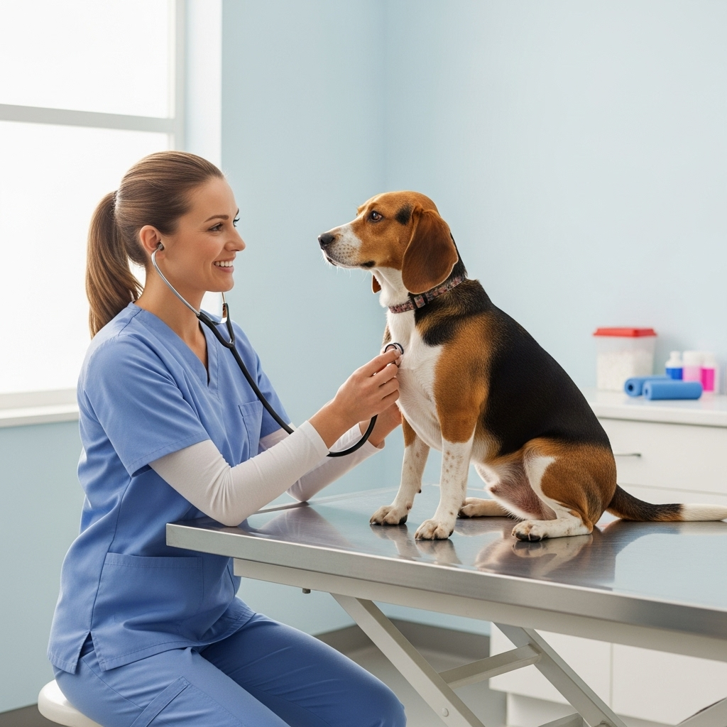
- It starts with a Conversation and Exam: Your observations are critical. A thorough nose-to-tail physical exam by your vet is the non-negotiable first step. This initial hands-on assessment provides the first clues.
- Imaging Looks Deeper: We use tools like X-rays and ultrasounds to see what’s happening inside your dog’s body. These tests help determine the size and location of a potential tumour and, crucially, check if it has spread to other areas like the lungs.
- A Biopsy is the Only Way to Be Sure: While imaging is vital, only a tissue sample (a biopsy) can provide a definitive cancer diagnosis. This tells us exactly what type of cells we’re dealing with, which is the gold standard for creating an effective treatment plan. The entire process of diagnosing cancer in dogs hinges on the information a biopsy provides.
The First Step: What Happens During the Initial Vet Visit?
You can Also Visit: https://doglifeexpert.com/heartworm-prevention-for-dogs/
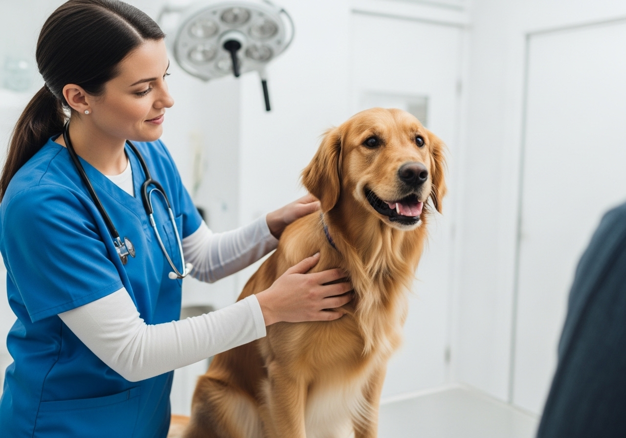
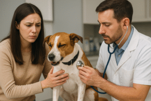
The journey of diagnosing cancer in dogs always begins in the quiet of the exam room. It’s a partnership from the very start. Your role is just as important as mine.
The Nose-to-Tail Physical Exam: More Than Just a Pat-Down
Before we reach for any advanced tools, our most sensitive and effective diagnostic instruments are our hands and eyes. A thorough physical exam provides a wealth of information.
I remember a sweet senior Labrador named Max who came into my Ottawa clinic. His owner had found a small, soft lump on his side. During the exam, I carefully felt the lump, noting its size, whether it was attached to the underlying muscle, and if it was painful. But I didn’t stop there. I checked his lymph nodes (they were slightly enlarged), listened to his heart and lungs, and gently palpated his abdomen. This complete assessment is fundamental. A single lump can sometimes be a sign of a more systemic issue, and a comprehensive exam helps us build the full picture. This hands-on approach is the cornerstone of diagnosing cancer in dogs.
Your Crucial Role: Discussing Symptoms and History
You see your dog every day. You know what’s normal and what’s not. The information you provide is invaluable. I’ll ask questions like:
- When did you first notice the lump or symptom?
- Has it changed in size, shape, or texture?
- Have you noticed any changes in appetite, energy levels, or water consumption?
- Is there any coughing, vomiting, or change in bathroom habits?
Your detailed answers guide the entire process of diagnosing cancer in dogs. What might seem like a minor detail to you could be a critical clue for me.
Seeing the Unseen: Core Imaging Tests for Diagnosing Cancer in Dogs
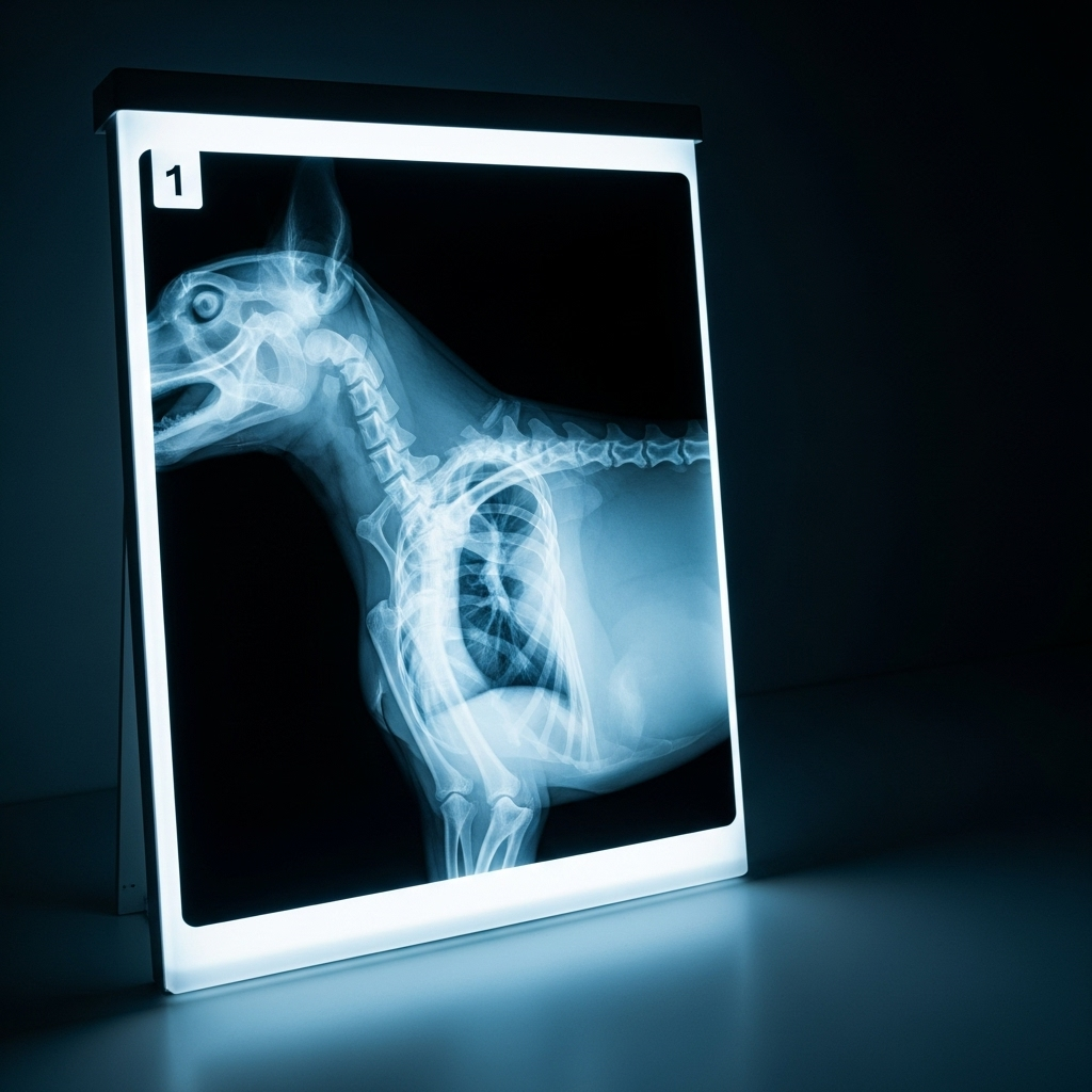
Once the physical exam points us in a specific direction, we need to look inside the body. Diagnostic imaging allows us to visualize structures we can’t see or feel, which is a critical part of diagnosing cancer in dogs.
X-Rays: The Foundational First Look
Radiographs, or X-rays, are often our first imaging step. They are excellent for evaluating bones and are particularly vital for checking the chest. One of our biggest concerns with any lump is whether it has metastasized, or spread. The lungs are a common site for metastasis, so taking chest X-rays is a standard procedure when staging a potential cancer. This helps us understand the scope of the problem we might be facing.
Ultrasound: A Deeper, Real-Time View
If we need to examine soft tissues, an ultrasound is the tool of choice. It uses sound waves to create a real-time image of abdominal organs like the spleen, liver, and kidneys. If I felt something abnormal during Max’s abdominal palpation, an ultrasound would be my next recommendation. It allows us to see the internal architecture of an organ, identify masses, and even guide a needle for a biopsy. It provides a level of detail that X-rays simply can’t for soft tissues, making it a key procedure for diagnosing cancer in dogs.
Advanced Imaging: When CT and MRI are Necessary
For complex cases, especially those involving the brain, nasal passages, or intricate bone tumours, we may need more advanced imaging.
- Computed Tomography (CT): A CT scan is like a three-dimensional X-ray. It takes multiple images in “slices” and compiles them into a highly detailed 3D model. This is invaluable for surgical planning, as it shows the exact extent of a tumour and its relationship with surrounding tissues.
- Magnetic Resonance Imaging (MRI): An MRI uses powerful magnets and radio waves to create incredibly detailed images of soft tissues, particularly the brain and spinal cord. It’s the gold standard for diagnosing brain tumours.
While not every case requires these advanced tools, they are essential for getting the clearest possible picture when the stakes are high in diagnosing cancer in dogs.
The Definitive Answer: Why a Biopsy is the Gold Standard
I often tell clients, “We can suspect with imaging, but we can only diagnose with a biopsy.” This is the most critical concept in veterinary oncology. No matter what an X-ray or ultrasound shows, a tissue sample is the only way to get a definitive answer. The entire approach to diagnosing cancer in dogs leads to this pivotal step.
Fine-Needle Aspirate (FNA): The Least Invasive Step
An FNA is often the first type of biopsy we’ll perform. It involves using a small needle, similar to a vaccine needle, to collect a sample of cells from a lump. It’s quick, minimally invasive, and often doesn’t require sedation.
The collected cells are placed on a microscope slide and examined by a pathologist. This can often tell us if we’re dealing with inflammation, a benign growth (like a fatty lipoma), or something more concerning. However, an FNA only collects a small number of cells, so sometimes the results are inconclusive. It’s a great screening tool in the process of diagnosing cancer in dogs.
Surgical Biopsy: Getting the Full Picture
For a definitive diagnosis and to determine the grade of a tumour (how aggressive it is), a larger tissue sample is needed. This is done through a surgical biopsy, which typically requires heavy sedation or general anesthesia.
There are two main types:
- Incisional Biopsy: A small piece of the tumour is removed. This is done when the tumour is very large or in a difficult location, and we need a diagnosis before planning a major removal surgery.
- Excisional Biopsy: The entire lump is removed with clean margins of healthy tissue around it. This is both diagnostic and, in many cases, curative.
The tissue is then sent to a lab where a pathologist performs a histopathological evaluation. This report is the roadmap for treatment. It tells us the type of cancer, the grade, and whether the margins are “clean,” indicating the entire tumour was likely removed. This detailed information is why a biopsy is considered essential for diagnosing cancer in dogs. As outlined in foundational texts like Blackwell’s Five-Minute Veterinary Consult and the Saunders Manual of Small Animal Practice, obtaining a definitive histologic diagnosis is paramount before embarking on any treatment plan.
The Future is Here: Emerging Biomarkers in Canine Cancer Diagnosis
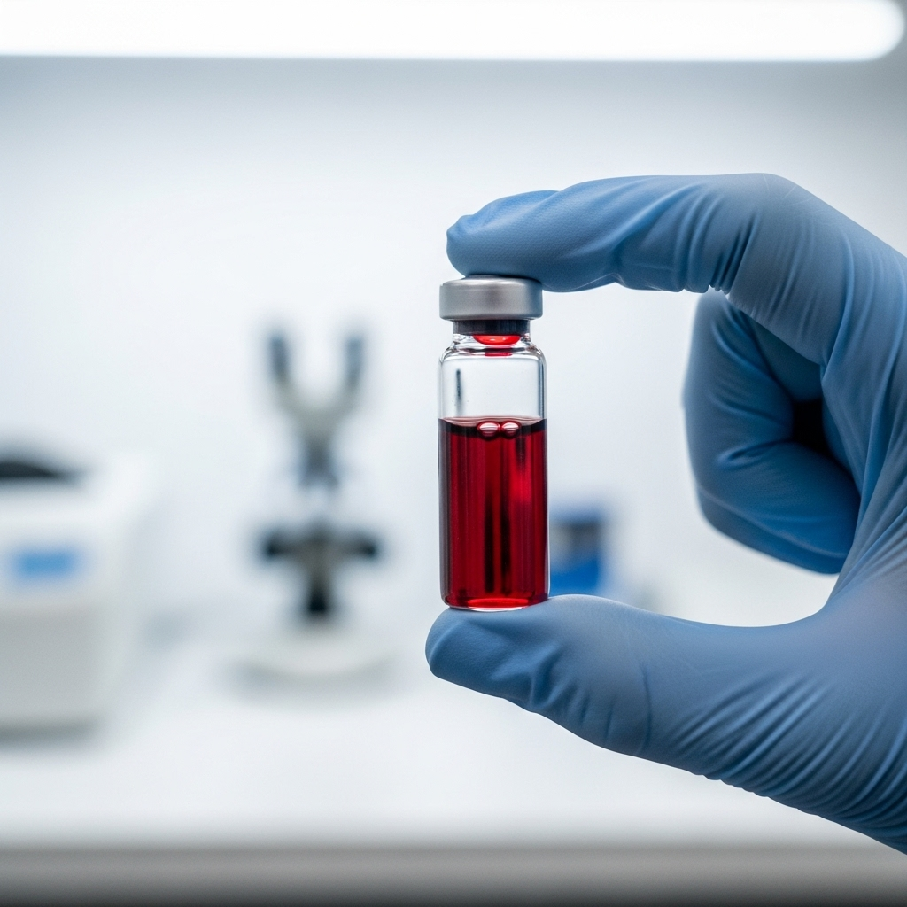
The field of veterinary oncology is constantly advancing. While physical exams, imaging, and biopsies remain the core of diagnosing cancer in dogs, we are on the cusp of exciting new technologies. One of the most promising areas is the use of biomarkers, particularly through “liquid biopsies.”
A liquid biopsy is a blood test that can detect cancer DNA circulating in the bloodstream. This technology is still emerging, but it holds incredible potential. A 2023 study in the Journal of the American Veterinary Medical Association explored the clinical use of these tests, highlighting their potential to help with early detection and monitoring treatment response. While not yet a replacement for traditional methods, it represents a significant leap forward in how we may soon be diagnosing cancer in dogs.
These advancements are part of a broader evolution in veterinary medicine. As researchers gain new insights into veterinary cancer immunology, our ability to diagnose and treat cancer with greater precision will only grow. This focus on the molecular level of disease, as detailed in resources like the Merck Veterinary Manual‘s overview of cancer treatment principles, is the future of care.
Actionable Checklist: A 5-Minute At-Home Health Check for Your Dog

Regularly checking your dog at home can help you spot potential problems early. Set aside five minutes each month to perform this gentle check.
- Nose-to-Tail Palpation: Gently run your hands over every part of your dog’s body, including their head, neck, chest, back, legs, and tail. Feel for any new lumps, bumps, or areas of swelling.
- Check the Mouth: Lift their lips and look at their gums and tongue. Note any unusual bumps, discoloration, or bad odours.
- Monitor Energy & Appetite: Have their energy levels dropped significantly? Are they suddenly picky eaters, or have they lost weight unexpectedly?
- Watch Bathroom Habits: Keep an eye out for any straining to urinate or defecate, or any blood in their urine or stool.
- Note Behavioural Changes: Is your dog hiding more, showing signs of pain, or having difficulty walking?
These are some of the key warning signs of cancer in pets, as outlined by the Veterinary Cancer Society. If you notice any of these signs, it’s time to book a visit with your vet for a professional evaluation, which is the first step in diagnosing cancer in dogs.
Understanding the Costs: A Realistic Look at Diagnosing Cancer in Dogs in Canada
The cost of diagnosing cancer in dogs can be a major concern for pet owners. Being prepared can help you make decisions without financial pressure clouding your judgment. Costs can vary significantly based on your location in Canada and the specific clinic, but here is a general guideline.
| Diagnostic Procedure | Typical Cost Range (CAD) | Purpose |
| Initial Consultation & Exam | $80 – $150 | The essential first step for any health concern. |
| Chest X-Rays (2-3 views) | $250 – $450 | To check for metastasis (spread) to the lungs. |
| Abdominal Ultrasound | $400 – $700 | To examine soft tissue organs like the spleen and liver. |
| Fine-Needle Aspirate (FNA) | $100 – $250 | A minimally invasive cell sample for initial screening. |
| Surgical Biopsy (inc. anesthesia & pathology) | $800 – $2,500+ | The gold standard for a definitive diagnosis; price varies with complexity. |
These figures represent the diagnostic phase only. The costs of treatment, should a cancer diagnosis be confirmed, are separate.
Frequently Asked Questions about Diagnosing Cancer in Dogs
How can I tell if my dog’s lump is cancerous at home?
Unfortunately, you can’t. There is no way to know for sure if a lump is cancerous just by looking at it or feeling it. Some benign lumps can feel firm and irregular, while some aggressive cancers can feel soft and movable. The only way to know for certain is to have a veterinarian perform a biopsy. This is the most crucial part of diagnosing cancer in dogs.
Can blood tests alone detect cancer in dogs?
Generally, no. Standard blood work (like a complete blood count and chemistry panel) can give us clues about a dog’s overall health and may show abnormalities associated with certain cancers, but it cannot diagnose cancer on its own. The emerging “liquid biopsy” blood tests are a promising future tool for diagnosing cancer in dogs, but they are not yet a routine part of the initial diagnostic process.
How long does it take to get biopsy results for a dog?
The timeline can vary. For a fine-needle aspirate (FNA) that is read by an in-house pathologist, you might get results within a day. For a surgical biopsy that is sent to an external laboratory, it typically takes 5 to 10 business days to get the full, detailed histopathology report. This wait can feel long, but the comprehensive information is vital for proper treatment planning.
What are the 10 most common warning signs of cancer in dogs?
The Veterinary Cancer Society lists ten key warning signs to watch for: abnormal swellings that persist or grow, sores that don’t heal, weight loss, loss of appetite, bleeding or discharge from any body opening, offensive odour, difficulty eating or swallowing, hesitation to exercise or loss of stamina, persistent lameness or stiffness, and difficulty breathing, urinating, or defecating. Seeing any of these should prompt a veterinary visit.
A Concluding Thought: Your Partner in Your Dog’s Health
Facing the possibility of cancer in your beloved companion is one of the hardest things a pet owner can go through. The process of diagnosing cancer in dogs, with its exams, tests, and waiting, can feel overwhelming. But you are not alone in this.
Think of your veterinarian as your dedicated partner, here to navigate every step with you. We are here to provide answers, offer options, and support you with compassion and expertise. The journey begins with a single conversation. By working together, we can face the uncertainty head-on and make the best possible decisions for the friend who depends on you completely. Your watchfulness at home and our clinical tools are a powerful combination, ensuring your dog gets the care they deserve, right when they need it most.


Pingback: Blood Tests for Dogs: 3 Essential Screens Your Vet Recommends